- Introduction to Subjective Methods
- Birth weight
- Body shape
- Weight and height
- Waist and hip circumference
- Introduction to Objective Methods
- Simple measures - stature
- Simple measures - weight
- Simple measures - circumference
- Simple measures - arm anthropometry
- Simple measures - skinfolds
- Simple measures - abdominal sagittal diameter
- Simple measures - head circumference
- Bioelectric impedance analysis
- Multi-component models
- Hydrostatic underwater weighing
- Air displacement plethysmography
- Hydrometry
- Whole body DEXA scan
- Near infrared interactance
- Whole body counting of total body potassium
- 3d photonic scan
- Magnetic resonance imaging (MRI) / Magnetic resonance spectroscopy (MRS)
- Total body electrical conductivity (TOBEC)
- Computed tomography (CT)
- Ultrasonography
- Introduction anthropometric indices
- Body mass index
- Fat and fat free mass indices
- Ponderal index
- Percentiles and Z-scores
- Anthropometry Video Resources
- Height procedure
- Protocol for measuring waist circumference
- Measuring hip circumference
- Weight and body composition procedure
3d photonic scan
Three-dimensional photonic scanning (3D-PS) captures body surface topography, from which extensive body shape and anthropometrics can be extracted using computer algorithms.
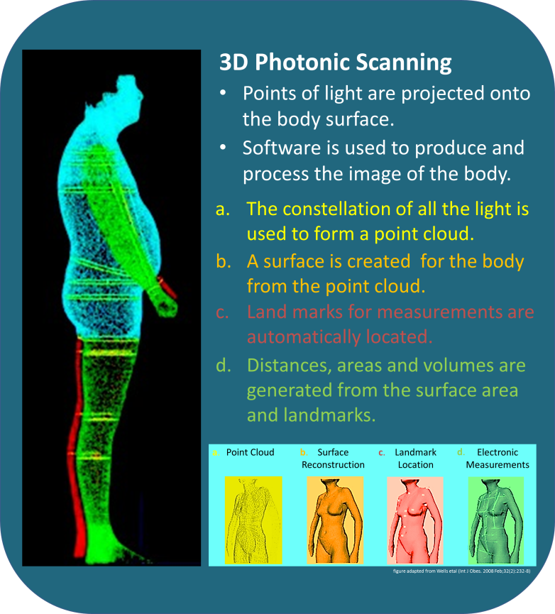
This technique projects light (typically infrared) on to the body surface, and cameras record the distortion of these light patterns to generate a point cloud. Computer algorithms then reconstruct the 3D body surface topography (see Figure 1), allowing automatic landmarks to be located through customised software. From these landmarks, a variety of girths, diameters and volumes can be derived using further algorithms.
The protocol for conducting three-dimensional photonic scan involves the following stages:
- The participant wears form-fitting clothing and adopts a standardised position for the scan. The scan can be initiated by either the participant or operator depending on device and protocol.
- The scanners project onto the body form and records the distortions induced by the body’s surface topography. During the scan participants must stand motionless while holding their breath, which is done immediately after a maximum expiration.
- The scanner generates a 3D point cloud (75 points per cm², device dependent) which represents a constellation of points located on the body’s surface in three dimensions.
- The specialised software generates a body surface and locates key landmarks on the digital body, such as the shoulders, breasts, navel, and so on.
- From these landmarks, measurements such as those for the bust, waist, and hips can be generated. This is similar to placing a virtual tape measure around different parts of the body and reading off the measurements. Figure 1 shows the different stages from point cloud to virtual tape measure.

Figure 1 Image processing involves the application of computer algorithms to clean (a) the point cloud to reconstruct (b) the skin surface. The software then automatically identifies (c) body landmarks, allowing (d) application of an e-tape measure,
generating digital information on distances, girths and curvature.
Adpated from: [7].
Three dimensional photonic scanning is a feasible alternative to traditional anthropometry. It is much quicker, precise and provides additional relevant information (e.g. shape and size variability). It has the potential to contribute in a number of ways to research and to clinical practice.
The success of several large national sizing surveys in the UK and US demonstrate the capacity of the technique to be applied to large numbers of individuals across the entire adult age range, and across the spectrum of ethnic variability.
The ease of use, high acceptability and low time requirement per participant makes 3D-PS ideal for large-scale anthropometric surveys, potentially including obesity screening. Furthermore, the visualisation provided by this technique is likely to aid/maintain patient motivation and hence compliance, a key issue in obesity management given the tendency for individuals to regain weight previously lost.
The method does not assess body composition values per se, but it provides an array of measurements that capture body size and shape.
The system used determines the density of the point cloud and the software used for the generation of estimates as they are typically integrated. Data processing and analysis is conducted by proprietary software. Initial data processing cleans the point cloud by rejecting inconsistent data points arising from artifacts in the scanning area. Software incorporates automatic landmark identification, whereby algorithms automatically extract key anatomical locations. These are then used to guide an ‘e-tape measure’, which provides a large number of girths, depths and diameters.
Using the different dimensions extracted from the scanner, principal component analysis (PCA) can be used to investigate the variation of body shape in the data set. Where the data does not assume a normal distribution, independent component analysis can identify more distinct (or independent) modes of variation in body shape.
Key characteristics of three-dimensional photonic scanning are summarised in Table 1.
Strengths
- Low cost per participant. In addition, the cost per photonic instrument has declined rapidly as commercial uptake of the technology has increased. Whole body photonic scanners could be installed in clinics, hospitals and health centres for routine clinical monitoring of body shape and size.
- Ease of use.
- It captures body shape and size variation.
- It may identify other anthropometric components which traditional anthropometry or body composition assessment methods do not capture.
- High acceptability to participant.
- Low time requirement - raw data collection is extremely rapid, lasting only a few seconds
- Provides visualisation of data which can aid compliance.
- A wide variety of digital shape outputs can be extracted.
- Scans can be electronically archived for analysis with improved software in the future.
- Digital outputs can extend to 2D or 3D format, whereas manual measurements (e.g. girths) are only 1D.
Limitations
- Movement artefacts may affect validity.
- It has not been validated in many populations.
- The validity of the e-tape measures to other established instruments is not clear.
- The clinical value of these measurements is still under research.
- There are no equations available to determine body composition (e.g. body fat).
Table 1 Characteristics of three dimensional photonic scanning.
| Consideration | Comment |
|---|---|
| Number of participants | Large |
| Relative cost | Medium |
| Participant burden | Low |
| Researcher burden of data collection | Low |
| Researcher burden of coding and data analysis | Low |
| Risk of reactivity bias | No |
| Risk of recall bias | No |
| Risk of social desirability bias | No |
| Risk of observer bias | Yes |
| Space required | Medium |
| Availability | Medium |
| Suitability for field use | Not suitable |
| Participant literacy required | No |
| Cognitively demanding | No |
Considerations relating to the use of 3D-PS for anthropometry in specific populations are described in Table 2.
Table 2 Anthropometry by 3D-PS in different populations.
| Population | Comment |
|---|---|
| Pregnancy | Suitable in principle. However, it requires validation. |
| Infancy and lactation | May not be suitable as the protocol requires the participant to stay still for 1- 24 seconds (depending on the instrument used). |
| Toddlers and young children | 3D-PS is acceptable in children aged 5 years and above, though with current hardware/software, and body movement artefacts, approximately one third of scans may be unsuccessful because of movement. |
| Adolescents | Suitable |
| Adults | Suitable |
| Older Adults | Suitable as long as participant is able to stand still e.g. may not be applicable in Alzheimer patients or in individuals with neurological disorders that can produce tremor. |
| Ethnic groups | Suitable |
| Other (obesity) | Suitable |
Refer to section: practical considerations for objective anthropometry
A method specific instrument library is being developed for this section. In the meantime, please refer to the overall instrument library page by clicking here to open in a new page.
- Lessa Horte B, Gigante DP, Goncalves H, dos Santos Motta JV, de Mola CL, Oliveira IO, Barros FC, Victora CG: Cohort Profile Update: The 1982 Pelotas (Brazil) Birth Cohort Study Int J Epidemiol 2015: 1
- Milanese C, Giachetti A, Cavedon V, Piscitelli F, Zancanaro C: Digital three-dimensional anthropometry detection of exercise-induced fat mass reduction in obese women Sports Sci Health 2015: 11; 67
- Santos LP, Ong KK, Day F, Wells JCK, Matijasevich A, Santos IS, Victora CG, Barros AJ: Body shape and size in 6-year old children: assessment by three-dimensional photonic scanning Int J Obesity 2016: 40; 1012
- Wang J, Gallagher D, Thornton JC, Yu W, Horlick M, Pi-Sunyer FX: Validation of a 3-dimensional photonic scanner for the measurement of body volumes, dimensions, and percentage body fat Am J Clin Nutr 2006: 83; 80
- Wells JCK, Treleaven P, Charoensiriwath S: Body shape by 3-D photonic scanning in Thai and UK adults: comparison of national sizing surveys Int J Obesity 2012: 36; 148
- Wells JCK, Stocks J, Bonner R, Raywood E, Legg S, Lee S, Treleaven P, Lum S: Acceptability, Precision and Accuracy of 3D Photonic Scanning for Measurement of Body Shape in a Multi-Ethnic Sample of Children Aged 5-11 Years: The SLIC Study PLoS ONE 2015: 10; e0124
- Wells JCK, Ruto A, Treleaven P: Whole-body three-dimensional photonic scanning: a new technique for obesity research and clinical practice Int J Obesity 2008: 32; 232
